The Science Behind X-Rays Explained
It seems strange to think that things can move through your body without you being aware of it, but that's what happens when you get an X-ray. Invented in 1895, X-rays came about mostly as an accident when German physicist Wilhelm Conrad Röntgen was experimenting with cathode rays. According to History, he was curious to know if the rays could pass through glass and, through a series of experiments, found out that they caused a chemically coated screen to glow. Not only that, but they appeared to be able to move through not only glass but other things like human flesh. However, they didn't pass through other things, such as bone.
So how is it that rays pass through your body? The answer, without getting too complicated, has to do with the kind of energy X-rays are made of — specifically, electromagnetic energy, or photons. X-rays are like light rays, but much shorter, which is why we can't see them, per Interesting Engineering.
X-rays push electrons through the air

In order to take an X-ray, you need a machine that contains an X-ray source and an X-ray detector, according to the National Institute of Biomedical Imaging and Bioengineering (NIBIB). When the machine is turned on, it creates a reaction that releases the electromagnetic energy and pushes it through the air at a high velocity.
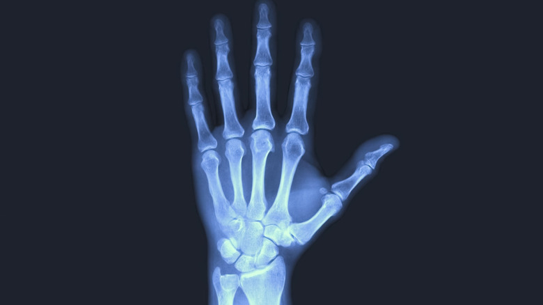
When it comes to taking an X-ray of the human body, doctors use film that is sensitive to X-rays and place it somewhere on the other side of the body where it can capture an image of the energy. Interesting Engineering explains that sometimes, doctors inject liquid contrast to look at blood vessels. Doctors also use fluoroscopes when they are looking at soft tissue, such as muscle.
There is no doubt Röntgen's discovery changed medicine, but it took some time to figure out that X-rays produced harmful radiation that could damage living tissue. If X-rays are used appropriately, and in low doses, they are safe, per History. That said, modern medicine offers many alternatives to X-rays, such as an MRI (magnetic resonance imaging) or ultrasound, per NIBIB.